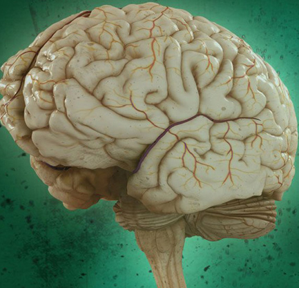
A decade ago, the Human Brain Project launched with a blue-sky goal: digitizing a human brain. The implications were huge: simulated brains could provide crucial clues to help crack some of the most troubling neurological diseases.
Rather than using animal models, they might better represent an Alzheimer’s brain, or one from people with autism or epilepsy. In a study published this January, the teams showed that virtual brain models of people with epilepsy can help neurosurgeons better hunt down the brain regions responsible for their seizures.
Each virtual brain tapped into a computational model dubbed the Virtual Epileptic Patient, which uses a person’s brain scans to create their digital twin.
With a dose of AI, the team simulated how seizure activity spreads across the brain, making it easier to spot hotspots and better target surgical intervention.
The method is now being tested in an ongoing clinical trial called EPINOV. If successful, it’ll be the first personalized brain modeling method used for epilepsy surgery and could pave the road for tackling other neurological disorders.
The results will be part of the legacy of the Virtual Brain, a computational platform to digitize personalized neural connections.
To be clear: the models aren’t exact replicas of a human brain.
“As evidence accumulates in support of the predictive power of personalized virtual brain models, and as methods are tested in clinical trials, virtual brains might inform clinical practice in the near future,” Jirsa and colleagues wrote.
Large-scale brain mapping projects now seem trivial. Neuroscientists were hunting down the neural code-the brain’s algorithms-with success, but in independent labs.
With more than 500 scientists across 140 universities and other research institutions, the European Union project became one of the first large-scale programs-along with the US’s BRAIN Initiative and Japan’s Brain/MINDS-to attempt to solve the brain’s mysteries by digitally mapping its intricate connections.
In turn, it’s hoped, the global effort can generate better models of the brain’s inner workings.
Why care? Our thoughts, memories, and emotions are all encoded in the brain’s neural networks.
Like how Google Maps for local roads gives insight into traffic patterns, brain maps can spark ideas on how neural networks normally communicate-and when they go awry.
Epilepsy affects roughly 50 million people worldwide and is triggered by abnormal brain activity.
The brain’s electrical activity “Hums” at different frequencies.
Like a pair of basic headphones, SEEG captures high-frequency brain activity but misses the “Bass”-low-frequency aberrations sometimes seen in seizures. In the new study, the team integrated all these test results into the Virtual Epileptic Patient model built on the Virtual Brain platform.
It starts with images of each patient’s brain from MRI and CT scans-the latter track down the white matter highways connecting brain regions.
In a retrospective test of 53 people with epilepsy, they used these virtual brains to hunt down the brain region responsible for each person’s seizures by triggering seizure-like activity in the digital brains.
The team generated a virtual brain for a patient who had 19 parts of his brain removed to rid him of his seizures.
They’re personalized atlases of 162 brain regions with a resolution of around one square millimeter-roughly the size of a small grain of sand.
Scientists will follow up on their outcomes for a year to see if a digital surrogate brain helps keep them free of seizures.
Despite a decade of work, it’s still early days for using virtual brain models to treat disorders.
“Computational neuromedicine needs to integrate high-resolution brain data and patient specificity,” he said.
“Our approach heavily relies on the research technologies in EBRAINS and could only have been possible in a large-scale, collaborative project such as the Human Brain Project.”
