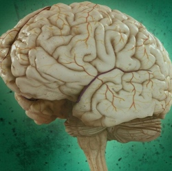
Using a new high-speed, high-resolution method, researchers were able to see blood flow, blood oxygenation, oxygen metabolism, and other functions inside a living mouse brain at faster rates than ever before. Researchers rely on high-resolution imaging to see tumors and other activity deep within the body’s tissues.
The new method is called “photoacoustic microscopy” (PAM), a single-wavelength, pulse-width-based technique developed by Lihong Wang, PhD, the Gene K. Beare Professor of Biomedical Engineering in the School of Engineering & Applied Science, and his team.
PAM allows for capturing images of blood oxygenation 50 times faster than their previous results using fast-scanning PAM, reported on KurzweilAI in November. The new technology is also 100 times faster than their acoustic-resolution system, and more than 500 times faster than phosphorescence-lifetime-based two-photon microscopy (TPM).
Other existing methods, including functional MRI (fMRI), TPM and wide-field optical microscopy, have provided information about the structure, blood oxygenation and flow dynamics of the mouse brain. However, those methods have speed and resolution limits, Wang says.
To make up for these limitations, Wang and his lab implemented “fast-functional PAM,”which allowed them to get high-resolution, high-speed images of a living mouse brain through an intact skull. This method achieved a lateral spatial resolution five times finer than the lab’s previous fast-scanning system; 25 times finer than its previous acoustic-resolution system; and more than 35 times finer than ultrasound-array-based photoacoustic computed tomography.
Most importantly, PAM allowed 3-D blood oxygenation imaging with capillary-level resolution at a one-dimensional imaging rate of 100 kHz, or 10 microseconds.
“Using this new single-wavelength, pulse-width-based method, PAM is capable of high-speed imaging of the oxygen saturation of hemoglobin,” Wang said. “In addition, we were able to map the mouse brain oxygenation vessel by vessel using this method.”
